

Current trends in animal cell research show progress in creating viable 3D cell culture as a surrogate for animal testing. Animal testing, which has notably been used in many concepts historically such as Koch’s postulates and as a part of current drug manufacturing practices, is a prevalent method to test scientific ideas without testing on humans. However, the ethical issues, barriers and compatibility issues that come with animal testing causes people to frown upon this technique. Although 2D cell culture has been around for a while and used for things such as drug development, the major issue is that 2D cell cultures tend to lack that physiological similarity present in vivo. This is due to the dependence 2D cell cultures have on where they anchor (where they attach to). The main reason this makes drug research so difficult is because they react by anoikis (cell death caused by cell detachment). Anoikis may lead to false efficacy data as it would not truly simulate the same as drug induced apoptosis.
3D cell research circumvents these issues allowing for better drug treatments and in vivo analogues. Although 3D cell culture models have existed long term, this research shows promise as they do testing on NIDs (new investigational drugs) which is something that has not occurred in the past using a 3D cell culture model. The authors hypothesize that this model using MCTS (multicellular tumor spheroids a.k.a “giant 3D spere of cancer cells”) can be used to test new investigational drugs without the use of animals or 2D cells which don’t work. Thus the scientists do a comparative study between known cancer drugs and two new drugs to determine if MCTS are an efficient analogue. Alongside research with standard MCTS the scientists also experiment to see if MCTS produced via perfusion of cancer cells into scaffolds work as a better analogue than the standard MCTS.
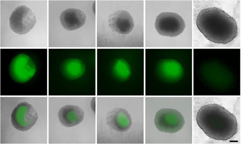
Bar, Biersack, and Schobert go about their research by culturing colon cancer cells and then creating standard MCTS, basically a sphere of made tumor cells, by adding them to the proper environment of agarose wells after the cells were suspended over a solution that helps create the spheres. This leads to the creation of MCTS which can simulate a better in vivo microenvironment while being in vitro procedure. After creation they measure the diameter and volume of MCTS by inverted transmission microscopy. This is followed by visualization of dead cells present within the MCTS via stain to see how many dead cancer cells are present when drugs are added. Similarly, as a step prior to the main part of the experiment they test for initiator enzyme (caspase 9) expression within the MCTS. An LDH (lactate dehydrogenase) assay is preformed to measure LDH content which is directly linked to substance induced tissue death. Fluorescence microscopy is also used to determine ROS (reactive oxygen species a.k.a oxygen molecules that react within cell to cause apoptosis) generation. They also generate in vitro microtumors in scaffolds which are made using a perfusion system which lines cells. The scaffold is basically somewhere for the microtumors to be held within so that it conforms to a desired shape within a proper environment. Basically, they had compared the studies of plate based MCTS and scaffold grown MCTS and were able to conclude that this was a better representative of the in vivo experimentation done by drugs.
From these biotechnological techniques they were able to determine that MCTS works as a model for drug testing. Testing drugs and comparing the effects between known and new drugs led to results showing effectiveness. Using fluorescence, they were able to identify if the cell deaths were genuinely due to effectiveness of the compounds rather than the substances present within the compounds. They had also analyzed activity within the cell in relation to cell death.
They had concluded from this information that there is now a better way to test drugs using micro tumors. A more in-depth analysis shows that microtumors built from scaffolds are even better than standard MCTS created in a well-plate setup. The research shows promise that there can be 3D cell assays as a method to test drugs and the study of the new drugs showed effectiveness comparable to clinically established drugs. These experiments added to knowledge and advancements by showing proof of viability of 3D MCTS.