Imagine if you were an athlete preparing for a competition, but suddenly got injured during practice after a bad fall- you feel a sharp pain, but think that it wasn’t a big deal. Just brush it off, and continue on – practice is more important. However, the injury is worse than you thought, and the pain behind your injury persists. You see a doctor, and the doctor breaks to you the terrible news that the nerves in your injured area are completely damaged. What do you do? You don’t want to miss out on the competition, but your injury is severe. Luckily, current research in biomedical engineering is studying potential methods and advancing strategies for the reconstruction of neural tissues.
The Research
Research in biomedical engineering is studying methods on how in-vitro scaffolds could advance strategies for curing injuries and stimulating neural regeneration. Specifically, to advance the field of neural tissue engineering, researchers have to investigate how fiber morphology influences neural cell behaviour, organization, and differentiation.
But how exactly do these scaffolds work? How are neural cells able to spatially arrange themselves while communicating effectively with each other?
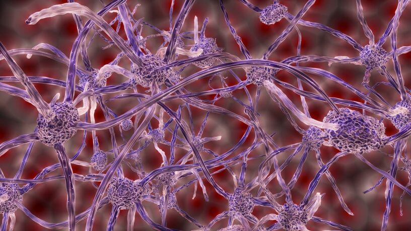
Fibrous scaffolds may be the key to answering this question – these scaffolds are extensively used in biomedical engineering research. The 3D structures of these fibrous scaffolds greatly resemble the extracellular matrix (ECM), which is a complex, woven network of proteins, cells, and molecules that provide structural support to cells. Therefore, fibrous scaffolds mimic a physiologically related environment for cell adhesion, growth, and networking. The 3D aspect of these scaffolds allows the cells to grow in different directions, which allows researchers to determine how the morphology of fibers influences growth, signaling, and the microenvironment of neural cells.
The Experiment and Crucial Findings

To investigate and understand fibrous scaffolds, researchers recreated them by monoaxial electrospinning polycaprolactone (PCL), which is a polymer used in biomedical engineering research. Electrospun PCL is crucial to this research as it is recognized for its ability to replicate the ECM and its function in providing structural support and biochemical cues for cellular differentiation and responses. Fibrous scaffolds in aligned and random orientations were produced before culturing human neuroblastoma cells SH-SY5Y on them. After culturing for one (D1) and seven (D7) days, cell viability, attachment, clustering, and gene expression were observed using Scanning Electron Microscopy (SEM) and confocal microscopy.
Fibers on the random and aligned scaffolds displayed distinct structural patterns and molecular responses, which not only indicated the importance of scaffold architecture but also supported the fact that fiber morphology and organization influence how cells grow, cluster, and mature. Although both cluster sizes and fiber coverage significantly increased over the seven days spent in incubation, fibers on the random scaffold were more expansive, whereas fibers on the aligned scaffold had deeper integration and distinct clustering dynamics rather than uniform spreading. The clustering dynamics observed in the aligned fibers indicated restricted cell-cell connectivity, which ultimately restricts cells from building an expansive network for communication. This is believed to have been caused by strong directional cues from the orientation of the scaffold.
Cells from the scaffold were cultured and measured via a series of experiments to gain data. Proteins and markers of the cytoskeleton, proliferation rates, fiber organization, and ECM from the fibers of the two scaffolds were compared and quantified from these experiments. Afterwards, confocal microscopy was used to analyze and image cells using immunofluorescence, while SEM micrographs were utilized to evaluate cluster sizes, morphology, migration, and coverage of cells.
In the first experiment, cytotoxicity assessment (LDH assay) and mitotic index were utilized to measure proliferation rates; however, no significant differences were found. Although the LDH assay displayed slightly lower values on fibers from the aligned scaffold, the results were not statistically significant. Therefore, surface morphologies are not expected to impact proliferation rates, but more research should be conducted before reaching a determined conclusion.
In another experiment, mRNA was taken from cells to undergo qPCR in order to determine gene expression of several structural and neural proteins.
The results show that mRNA expression for structural proteins has significant upregulation in random fibers, yet no upregulation in aligned fibers. Structural proteins such as actin and β3-tubulin play an important role in maintaining the cytoskeleton, hinting that higher cell migration and remodeling activity are occurring in random fibers.
Doublecortin (DCX) is a marker of neuronal identity, and was found to have a significant increase in aligned fibers compared to random fibers. An increase in DCX indicates that aligned fibers may be ideal for supporting new growth of neurons and promoting structural maturation.
Finally, Fibronectin 1 (FN1), a protein that influences cell adhesion, migration, and development in the ECM, displayed decreased expression in random fibers. The reduction of expression suggests increased migration, as cells that exhibited active cytoskeletal remodeling and migration had downregulation of FN1.
Implications and Conclusions
Using PCL to create two fibrous scaffolds of different morphology has allowed researchers to determine and understand how fiber morphology is influential in the growth, organization, and communication of neural cells. Various experiments, including LDH assay and qPCR, measured proteins and markers crucial to the neural regenerative process, demonstrating differences between fibers on aligned scaffolds and random scaffolds.
With these crucial implications, further research could be conducted to further advance the knowledge required for successful neuroregenerative treatments. One proposal for future experiments includes creating hybrid fibrous scaffolds, where random and aligned fiber regions could be combined and integrated. By doing so, a powerful scaffold with qualities such as rapid coverage seen in random fibers and directed nerve regrowth from aligned fibers can be achieved. With the acquired knowledge and future research, scaffold-based strategies can greatly advance in the field of neuroregenerative medicine.
Citations:
Hlinkova, J., Dziemidowicz, K., Ulrich, M. M., Agger, A. E., Lian, A-M., Reseland, J. E., & Samara, A. (2025). Neural growth patterns: how random and aligned fibers guide 3D cell organization and pseudospheroid formation. Frontiers in BIoengineering and Biotechnology, 13. doi: 10.3389/fbioe.2025.1659965
photo credits: pixabay
No AI was used in the writing of this article.
